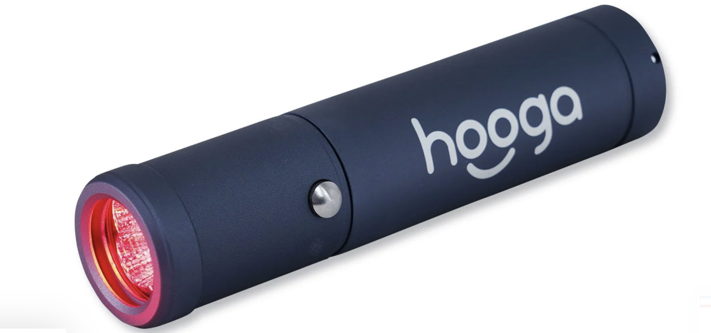
Hooga Red Light Therapy Torch Clinical Use Guide
Hooga Red Light Therapy Torch
Clinical Use Guide
Precision Tip • Focused Musculoskeletal & Tendon Applications
For clinician-to-clinician discussion and protocol development; not a manufacturer-validated treatment protocol.
Purpose & Clinical Rationale
The Hooga Torch is particularly well suited to localized, point-specific applications where the clinician can identify a small symptomatic tendon, enthesis, or fascial region. This guide focuses on plantar fascia pain, lateral epicondylitis, medial epicondylitis (golfer’s elbow), biceps tendon complaints, and patellar tendon complaints.
Photobiomodulation (PBM) should be viewed as an adjunct rather than a replacement for diagnosis, load management, progressive strengthening, mobility work, or other indicated rehabilitation. A 2021 systematic review/meta-analysis of 17 randomized trials (835 participants) found very-low-to-moderate quality evidence for PBM in tendinopathy; PBM combined with exercise showed improvements in some pain, function, and strength comparisons versus sham plus exercise, while results across interventions were heterogeneous.
Device Facts — Hooga Torch with Precision Tip
Parameter
Specification / Practical Note
Wavelengths:
630 & 660 nm red; 850 & 940 nm near-infrared; blue wavelength also available (manufacturer pages contain an inconsistency listing 415 vs 460 nm).
Irradiance:
Manufacturer reports approximately 120 mW/cm² at the surface.
Emitters:
5 high-output LEDs; rated 15 W, listed actual output 5 W.
Timer:
Built-in 3-minute session timer.
Precision tip:
Removable attachment for highly localized applications; remove it when a somewhat broader field is desired.
Manufacturer positioning:
Use just a little above the skin or target area.
Manufacturer session guidance:
1–3 minutes per area; daily use or as needed.
General Treatment Approach
For the musculoskeletal applications in this guide, use the red + near-infrared setting rather than blue-only light. Near-infrared wavelengths are particularly relevant when the target is beneath superficial skin layers.
Start conservatively and consistently. A practical starting range is 1–3 minutes at a clearly identified treatment point, consistent with Hooga’s device guidance. Do not assume that longer exposure is automatically better; PBM is dose-dependent and clinical studies use widely varying devices and dosing parameters.
Position the Torch just above the skin/target area as directed by Hooga. Avoid pressing the precision tip firmly into a painful tendon simply to get closer; mechanical compression can itself change pain and confound an immediate pre/post response.
When symptoms cover more than one small point, treat discrete clinically relevant points rather than repeatedly sweeping the device. The precision tip is most useful when the target is small; consider removing it when modestly broader coverage is desired.
Pair PBM with the condition-appropriate rehabilitation program. For tendinopathy, progressive loading remains the core intervention; PBM is best considered a potentially useful adjunct.
Condition-Specific Point Placement
Condition/Primary target/Starting application/Clinical note
Plantar fascia
Medial calcaneal tubercle / proximal plantar fascia at the primary symptomatic point.
1–3 min at primary point. If symptoms extend distally, consider a second discrete point along the symptomatic fascial band.
The fascia is superficial; pinpoint targeting is a strong fit for the precision tip.
Lateral epicondylitis
Common extensor tendon origin, often focused around the ECRB/common extensor region just distal/anterior to the lateral epicondyle.
1–3 min over the localized symptomatic tendon/enthesis. Add an adjacent point only if the symptomatic region clearly extends beyond the first target.
Avoid treating only the bony prominence when palpation/local loading identifies the tendon origin as the symptomatic tissue.
Medial epicondylitis / golfer’s elbow
Common flexor-pronator tendon origin at the medial epicondylar region.
1–3 min over the localized symptomatic tendon/enthesis.
Use anatomical care around the medial elbow; do not use the light application as a substitute for evaluating neural or other non-tendon sources of symptoms.
Proximal biceps tendon
Symptomatic proximal long-head biceps region/bicipital groove when clinically appropriate.
1–3 min over the localized target.
This target is deeper than the plantar fascia or epicondylar tendon origins; use red + NIR and interpret response cautiously.
Distal biceps tendon
Localized symptomatic distal tendon region when clinically appropriate.
1–3 min at the identified point.
Avoid unnecessary pressure over a painful tendon and reassess if symptoms suggest acute tearing or another condition requiring medical evaluation.
Patellar tendon
Most commonly the symptomatic proximal tendon/inferior pole region; alternatively tendon body or distal insertion if that is the clinically identified site.
1–3 min at the primary symptomatic point. Consider a second point if symptoms clearly span a longer segment.
The precision tip is useful for insertional/proximal pinpoint symptoms; larger or diffuse anterior-knee complaints may exceed the ideal use case of a small torch.
Dose & Frequency: Important Context
Hooga lists 1–3 minutes per target area and daily frequency for the Torch. Hooga’s broader educational material discusses approximate energy-dose ranges of 3–10 J/cm² for superficial/topical applications and 10–50 J/cm² for deeper tissues. Those general ranges should not be treated as a validated, tendon-specific prescription for this Torch: clinical PBM trials differ substantially in wavelength, irradiance, beam geometry, contact technique, dose, and treatment schedule.
Accordingly, it is preferable to standardize your own starting procedure (same mode, distance, site definition, and duration), track response, and avoid escalating exposure simply because a patient has not responded immediately.
Suggested In-Session Workflow
1. Identify the clinically relevant symptomatic structure and rule out reasons the complaint should not be treated as routine tendinopathy/fascial pain.
2. Record a reproducible baseline: pain rating and, when useful, a condition-specific provocative test (e.g., resisted wrist extension, heel loading, decline squat, tendon loading).
3. Identify and document the exact anatomical target point(s).
4. Select red + near-infrared mode for these musculoskeletal targets.
5. Position the device just above the target area; avoid firm pressure from the precision tip.
6. Treat the selected point for a conservative 1–3 minute session.
7. If a second point is clearly indicated, document and treat it separately rather than vaguely describing a large region.
8. Reassess the same baseline measure when clinically useful.
9. Continue the appropriate rehabilitation/loading program rather than using PBM as a stand-alone tendon intervention.
Documentation Template
Region / side
____________________________________________
Anatomical target
____________________________________________
Mode
Red + NIR
Duration / points
________ min × ______ point(s)
Pre-treatment response
Pain ____/10 | Provocative test: __________________
Post-treatment response
Pain ____/10 | Provocative test: __________________
Example: Right lateral elbow — ECRB/common extensor origin — Hooga Torch, red + NIR — 2 minutes — pain pre 5/10, post 3/10.
Precautions & Scope
Follow the manufacturer’s current manual and safety instructions. Hooga supplies protective eyewear and advises against use over the eyes or areas where light exposure may be unsafe.
Discontinue if the patient experiences irritation, unexpected discomfort, or an adverse response.
Exercise additional caution and follow manufacturer/medical guidance for patients with photosensitivity, photosensitizing medications, pregnancy, significant medical conditions, or other circumstances in which light therapy may be inappropriate.
Do not use PBM to delay evaluation of suspected rupture, fracture, infection, significant neurologic compromise, rapidly worsening symptoms, unexplained swelling, or another presentation requiring medical assessment.
The Hooga product information is marketed for general wellness and states that the device/information is not intended to diagnose, treat, cure, or prevent disease. Clinical use should remain within the practitioner’s professional scope, applicable regulations, and informed-consent procedures.
Key Takeaways for Our Use Case
The Torch’s strongest advantage is precision: it makes sense for the small, localized pain points described here.
Red + near-infrared is the practical mode for these tendon/fascial applications.
Use a standardized 1–3 minute starting application per discrete target point, just above the skin, rather than assuming more exposure is better.
Track exact site, time, mode, pre/post response, and any provocative test so repeated clinical experience becomes interpretable.
PBM should complement—not replace—progressive loading and condition-specific rehabilitation.
References
Hooga. Red Light Therapy Torch with Precision Tip — product specifications and session guidelines. Accessed September 7, 2026. https://hoogahealth.com/products/torch
Hooga. Red Light Therapy & PEMF FAQ — general dose guidance. Accessed September 7, 2026. https://hoogahealth.com/pages/faq
Hooga. Digital Resources / safety disclaimer. Accessed September 7, 2026. https://hoogahealth.com/pages/digital-resources
Tripodi N, Feehan J, Husaric M, et al. The effect of low-level red and near-infrared photobiomodulation on pain and function in tendinopathy: a systematic review and meta-analysis of randomized control trials. BMC Sports Sci Med Rehabil. 2021;13:91. doi:10.1186/s13102-021-00306-z.
Hooga Torch Clinical Use Guide • Prepared for clinician discussion • September 2026