EDGE Restriction System Blood Flow Restriction: Clinical Use Guide
EDGE Restriction System
BFR Clinical Use Guide
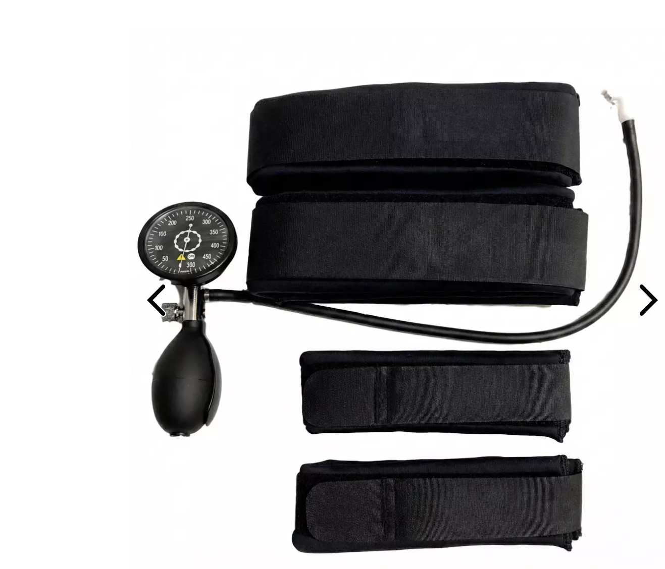
1 Arm Cuff + 1 Leg Cuff + Manual Pump/Gauge
Clinician reference for individualized blood-flow-restriction exercise and rehabilitation
For clinician-to-clinician discussion and protocol development; not a manufacturer-validated treatment protocol.
Purpose & Key Principle
Blood flow restriction (BFR) exercise uses an externally applied proximal limb cuff to partially restrict arterial inflow and substantially restrict venous outflow while exercise is performed at relatively low loads. It is not intended to create complete arterial occlusion during exercise.
The major clinical advantage is the ability to create a meaningful muscular training stimulus with loads that may be better tolerated when conventional high-load resistance is temporarily undesirable. BFR should complement—not replace—appropriate diagnosis, progressive loading, strength training, and condition-specific rehabilitation.
Your EDGE Equipment
Component/Manufacturer specification / practical point
Arm cuff
17¼ in long × 2 in wide.
Leg cuff
29¼ in long × 3 in wide.
Design
Single-bladder cuff with D-ring; cuffs are listed as waterproof and handmade in the USA.
Pump/gauge
Manual pump with quick-release valve; gauge reads to 500 mmHg.
Critical point
The gauge displays cuff pressure. It does NOT automatically determine an individual patient's limb occlusion pressure (LOP).
Manufacturer emphasis
EDGE specifically states that BFR exercise should use restriction rather than full occlusion.
Cuff Placement
Upper extremity: place the arm cuff as proximally as practical on the upper arm, near the axillary region. Do not place the BFR cuff around the forearm or directly over the painful elbow/tendon.
Lower extremity: place the leg cuff high on the proximal thigh near the inguinal/gluteal fold. Do not place the cuff around the knee, calf, patellar tendon, or painful structure.
The cuff creates the vascular restriction proximally; the exercise can target muscles and tissues distal to the cuff.
Pressure: Individualize With Limb Occlusion Pressure (LOP)
Avoid choosing the same arbitrary mmHg for every patient. Cuff width, limb circumference, tissue composition, blood pressure, cuff design, body position, and the individual all influence the pressure needed to occlude arterial flow.
Preferred approach: determine LOP with an appropriate objective method (commonly Doppler ultrasound; EDGE also teaches Doppler-based LOP determination). LOP is the minimum cuff pressure at which the distal arterial pulse is no longer detectable under the measurement conditions.
Then deflate and prescribe training pressure as a percentage of that measured LOP rather than exercising at 100% LOP. Common evidence-based implementation ranges are approximately 40–50% LOP for the upper extremity and 60–80% LOP for the lower extremity. Start toward the lower end when appropriate and individualize to tolerance, goals, patient characteristics, and your training/certification.
Reassess LOP when clinically appropriate, especially if cuff, limb, body position, or relevant patient factors change. Do not treat the pump's mmHg reading itself as a universal prescription.
Classic Low-Load BFR Resistance Protocol
Variable/Common starting framework
Load
~20–30% 1RM is a commonly used low-load range.
Sets / repetitions
30 reps → 15 → 15 → 15 (75 total repetitions), when appropriate.
Rest
~30 seconds between sets.
Cuff during set rests
Often remains inflated through the short inter-set rests in a standard resistance bout.
Between exercises / prolonged rest
Deflate as clinically appropriate; avoid unnecessary prolonged continuous restriction.
Frequency
Often 2–3 sessions/week in conventional programming; frequency should reflect tissue tolerance, rehab phase, total workload, and patient factors.
Failure
The goal is not automatically maximal failure on every set. Technique, symptoms, prescribed effort, and the rehab objective take priority.
Practical Applications With One Arm + One Leg Cuff
A single cuff is very useful for unilateral rehabilitation. Treat one limb, completely deflate/remove or transfer the cuff, then treat the other side if bilateral work is desired. Do not assume the pressure determined on one limb is automatically appropriate for the contralateral limb.
Clinical situation
How BFR can fit
Lateral / medial elbow tendinopathy
Arm cuff high on upper arm. Pair with appropriately loaded wrist extension/flexion, pronation/supination, grip, elbow or compound upper-extremity work according to the diagnosis and rehab phase.
Biceps-related rehabilitation
Arm cuff high on upper arm. Low-load elbow flexion and related upper-extremity exercise may allow a muscular stimulus with less external load. Respect tissue-healing restrictions after surgery or acute injury; BFR does not override surgeon/protocol loading limits.
Patellar tendon / anterior knee
Leg cuff high on thigh. Combine with tolerated quadriceps-focused exercise such as knee extension, squat/leg-press patterns, sit-to-stand or other appropriate kinetic-chain work.
Knee rehabilitation / quadriceps atrophy
Leg cuff high on thigh. BFR is especially relevant when heavier quadriceps loading is temporarily poorly tolerated or restricted. Exercise selection and ROM should follow the underlying diagnosis/procedure.
Calf / lower-leg strengthening
Leg cuff remains proximal on thigh; use appropriately selected calf or lower-extremity exercise. Do not move the cuff to the calf.
General deconditioning / return to loading
Use BFR as a bridge when low external loads are desirable, then progressively restore conventional loading as tolerated and indicated.
In-Session Workflow
1. Screen the patient and confirm that BFR is appropriate for the diagnosis, medical history, current status, and rehabilitation phase.
2. Explain expected sensations and obtain informed consent according to your clinical setting.
3. Record baseline symptoms and, when useful, a reproducible functional or strength measure.
4. Apply the correct cuff proximally on the limb.
5. Determine/document LOP with your chosen validated method under consistent conditions.
6. Select and document the prescribed percentage of LOP and resulting training pressure.
7. Choose exercise, load, ROM, sets/reps, rest, and effort appropriate to the patient's condition.
8. Monitor symptoms throughout the bout. Distal fatigue, muscular burning, pressure and effort are expected; alarming neurologic, vascular, systemic, or disproportionate symptoms are not.
9. Deflate promptly at completion or sooner if indicated; reassess the limb and patient.
10. Document pressure method, LOP, %LOP, exercise dose and response so future sessions are reproducible.
Stop / Deflate Immediately If
New numbness, tingling, loss of sensation, unusual weakness, or symptoms suggesting nerve compromise.
Severe or escalating pain that is not the expected muscular effort/burning of exercise.
Marked pallor, concerning discoloration, unusual coldness, or other signs suggesting compromised distal perfusion.
Dizziness, faintness, chest pain, unusual shortness of breath, severe headache, or other concerning systemic symptoms.
The cuff slips, pressure cannot be controlled, the equipment malfunctions, or you cannot appropriately monitor the patient.
Screening & Precautions
BFR is not appropriate for every patient. A formal pre-participation screening process is preferable to relying on a short universal contraindication list. Important issues that may warrant avoidance, medical clearance, or substantially greater caution include:
Known or suspected deep-vein thrombosis, pulmonary embolism, active clotting disorder, or significant thromboembolic risk.
Significant peripheral vascular/arterial disease, impaired circulation, vascular grafts or other relevant vascular pathology.
Uncontrolled or clinically significant hypertension or cardiovascular disease.
Pregnancy, depending on clinical context and medical guidance.
Active infection, significant open wound, or compromised tissue beneath/near cuff placement.
Significant neurologic impairment, altered sensation, inability to communicate symptoms reliably, or other circumstances that limit safe monitoring.
Recent surgery or acute injury where BFR has not been cleared or where the exercise/loading prescription conflicts with surgical precautions.
Any patient whose medical status, medications, history, or presentation raises concern for vascular, cardiovascular, neurologic, or thrombotic complications.
This list is intentionally not exhaustive. Use your professional scope, current evidence, formal BFR education, patient-specific risk assessment, and medical/surgical guidance.
Documentation Template
Region / side
____________________________
Cuff
Arm / Leg
LOP method
Doppler / other: ______________
Measured LOP
________ mmHg
Training pressure
________ % LOP = ________ mmHg
Exercise / load
____________________________
Sets / reps
____________________________
Rest / total restriction time
____________________________
Pre-treatment measure
____________________________
Post-treatment response / adverse signs
____________________________
Example Note
Right knee/quadriceps — EDGE leg cuff positioned proximal thigh — LOP measured with Doppler: ____ mmHg — training pressure ____% LOP / ____ mmHg — knee-extension exercise at prescribed low load — 30/15/15/15 with 30-sec inter-set rest — tolerated without neurologic/vascular warning symptoms — pre/post functional response documented.
Important Distinction: EDGE Instructions vs Clinical Programming
EDGE provides device specifications, emphasizes partial restriction rather than full occlusion, and offers education on determining LOP with Doppler. Exercise pressures, loading, repetitions, frequency, screening, and progression should be individualized using current evidence and clinician training rather than inferred solely from the pump gauge.
EDGE also offers a clinician manual and BFR certification resources. For routine patient use, formal BFR training is worth considering because the safety of the intervention depends heavily on screening, individualized pressure determination, cuff placement, exercise selection, and monitoring.
References / Resources
EDGE Mobility System. EDGE Restriction System BFR Cuffs — product specifications and manufacturer guidance. Accessed September 7, 2026.
EDGE Mobility System. The BFR Clinician's Manual — exercise prescription/programming resource; includes rehabilitation templates and Doppler use.
EDGE Mobility System. Edan Sonotrax Handheld Doppler Ultrasound — EDGE educational materials for finding upper- and lower-extremity pulses and limb occlusion pressure.
Patterson SD, Hughes L, Warmington S, et al. Blood Flow Restriction Exercise: Considerations of Methodology, Application, and Safety. Front Physiol. 2019;10:533.
Cognetti DJ, Sheean AJ, Owens JG. Blood Flow Restriction Therapy and Its Use for Rehabilitation and Return to Sport: Physiology, Application, and Guidelines for Implementation. Arthrosc Sports Med Rehabil. 2022;4(1):e71-e76.
Clinical disclaimer: This document is an educational clinician reference, not a substitute for BFR certification, manufacturer instructions, patient-specific medical evaluation, or applicable professional regulations.
EDGE BFR Clinical Use Guide • Clinician reference • September 2026